
Last updated: 7 November 2026
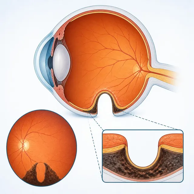
A retinochoroidal coloboma is a gap in the retina and the layer beneath it (the choroid) that is present from birth. It forms because a groove in the developing eye did not close completely during pregnancy. The gap usually sits in the lower part of the eye, which affects the upper visual field. Vision ranges from near normal to severely reduced, depending on how close the gap is to the macula and optic nerve.
Symptoms
Many people have no symptoms of their own, and the condition is found at a routine eye or school check. What you notice depends on size and position.
- Reduced vision in one or both eyes since childhood.
- A missing area in the upper field of vision (a dark patch above).
- A keyhole-shaped pupil, if the coloboma also involves the iris.
- Squint or nystagmus (shaky eye movements) in children.
Red flags
Flashes, a shower of floaters or a curtain of shadow need same-day review, because eyes with coloboma are more prone to retinal detachment. A child whose eyes do not follow light, or who has a white or keyhole-shaped pupil, should be examined soon.
Causes and risk factors
Coloboma arises when the optic fissure, a seam in the embryonic eye, fails to close in the early weeks of pregnancy. In many children no cause is found. Some are linked to gene changes and may be part of a syndrome that also affects the heart, ears, kidneys or growth (CHARGE syndrome is one example). Maternal infections or medicines in early pregnancy are other possible factors, though most cases cannot be traced.
How it is diagnosed
Diagnosis is by dilated fundus examination. The coloboma shows as a white or pale bowl-shaped patch with sharp edges, usually below the optic disc. Further tests help plan care:
- Vision and glasses power testing, with cycloplegic refraction in children.
- Fundus photography and OCT of the nearby retina and macula.
- B-scan ultrasound, if the eye is small or the view is poor.
- Visual field testing in older children and adults.
A paediatrician examines the child for associated conditions, and genetic counselling may be advised. Family members can be screened.
Treatment options
The gap itself cannot be closed or repaired, so treatment focuses on getting the best from the rest of the eye and preventing complications.
- Glasses and contact lenses: correct accompanying refractive error, which is common.
- Amblyopia treatment: patching or other therapy to build vision in a weaker child's eye. See squint and amblyopia .
- Regular retinal review: yearly, or as advised, to catch tears or thin areas early.
- Laser barrier: in selected eyes, laser around the edge may be considered to lower detachment risk, though evidence is limited and the decision is individual.
- Surgery: if the retina detaches, vitrectomy with gas or oil is used. These repairs are more complex than in an ordinary eye.
What Vision Plus does
Vision Plus offers dilated retinal evaluation, OCT, fundus imaging and retinal laser, with surgical theatres for retinal procedures, at Sector 51. Dr. Ajay Aurora manages retinal and vitreous aspects; Dr. Ritu Aurora handles children's glasses, amblyopia and squint. See Dr. Ajay Aurora , Dr. Ritu Aurora and retina, uvea and vitreous care . Families from Noida Extension can start at Gaur City-1.
Cost note
Cost depends on whether you need only check-ups and glasses or also laser, surgery or low vision aids. Please call the desk for current consultation fees. OCT is Rs 1,500 to 4,500. Final quote after examination. See eye treatment cost in Noida .
Living with it
Children with coloboma can learn and grow normally with the right support. Keep regular reviews, wear glasses as advised, use good lighting and large print, and tell the school. Protect the eyes from injury. Related pages: retinal detachment , paediatric eye diseases , and the eye diseases hub .
Frequently asked questions
What is a retinochoroidal coloboma?
It is a birth defect where part of the retina and choroid is missing, leaving a pale gap, usually in the lower eye. It is caused by incomplete closure of the eye's early groove. Vision depends on the size and position.
Is coloboma genetic?
Sometimes. Some cases are due to gene changes and may run in families or be part of a syndrome, while many have no clear cause. A paediatrician and genetic counselling help families understand recurrence and check other organs.
Can vision improve with treatment?
The missing tissue cannot be replaced, but glasses, amblyopia therapy and low vision aids can improve what the person can use. Results depend on whether the macula and optic nerve are healthy. No guaranteed outcome can be promised.
Does coloboma cause retinal detachment?
It raises the risk, so yearly retinal checks are advised. Report flashes, floaters or a shadow immediately. Detachment in these eyes is repairable in many cases but is more complex, and early treatment matters.
Where can I get a coloboma assessment in Noida?
Vision Plus Sector 51 or Gaur City-1. Call or WhatsApp +91 8800313134 to book a dilated retinal check, and bring any previous reports. You can also use the appointment page .
Book an examination
Call or WhatsApp +91 8800313134, or book at /appointment . Surgery consultation: +91 9717417417.
Sector 51: Kisan Tower, Capt Shashikant Sharma Marg (Mon to Sat 9:30 AM to 7:30 PM, Sun 10:00 AM to 4:00 PM). Gaur City-1: Aarza Square-2, Greater Noida West (Mon to Sat 10:00 AM to 7:00 PM, Sun closed).
Medically reviewed by Dr. Ajay Aurora, November 2026