
CORNEA - C3R, Keratoplasty
Recognised centre for Corneal transplants & treatment of corneal diseases
Have Any Query?
Enter Your Mobile Number For Call back
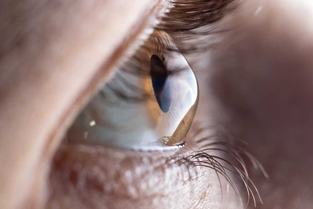
What is Cornea?
The Cornea is the transparent, outermost dome shaped part of the eye through which light enters the pupil and is focused on the retina. It is an extremely delicate structure,which requires normal functioning for accurate vision. Diseases and injuries of the cornea can permanently damage it, leading to severe vision loss. At Vision Plus Eye Centre we understand the importance of maintaining a healthy cornea and have an experienced team of Cornea Specialists backed by the latest diagnostic machines to give you the best cornea treatment in Noida & Delhi NCR. We treat all types of complex corneal disorders by both medical and surgical methods.
Understanding the Structure of the Cornea
- Normal cornea is 11.5mm in diameter & 520-540um thick.
- Functions such as positive meniscus lens- provides 2/3rd of the eye’s focussing power and hence we can correct refractive errors by changing its shape.
- Does not have blood vessels, gets oxygen and nutrients from the tear film outside and aqueous humour inside.
- Rich nerve supply which is highly pain sensitive
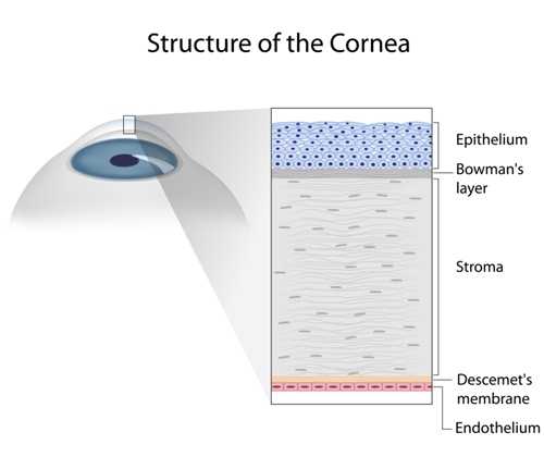
5 Layers of the Cornea
Epithelium Layer
The outermost layer which is about 50u in thickness and is made up of 6 to 8 layers of rapidly regenerating cells. It protects the eye from infection and foreign bodies and also absorbs nutrients and oxygen from the tear film which adheres to its shape. It is pain sensitive as it has a rich nerve supply If damaged (Corneal Abrasion) it heals by itself.
Bowman’s Layer
A tough layer of collagen that acts as protective. It can scar if injured.
Stroma Layer
Forms 90% thickness of the cornea. It is made of 78% water and 16% collagen fibrils that are arranged to give clarity. The stroma is reshaped during LASIK surgery. Adequate thickness of the stroma is essential for the normal strength and form. The keratocytes help in repairing if damaged.
Descemet’s membrane
Thin but strong layer of collagen which protects the eye and can regenerate after injury
Endothelium Layer
The innermost layer which is responsible for pumping out fluid and keeping the cornea transparent. A very low endothelial cell count can cause swelling and loss of vision. If damaged by injury, surgery or disease, Endothelial cells cannot regenerate.
Common Corneal disorders.
- Keratoconus
- Corneal Edema
- Corneal ulcer/ infection
- Pterygium
- Dry eye
- Corneal dystrophy
- Corneal scar/ opacity
- Corneal inflammation
- Ocular surface disorders
- Trauma

KERATOCONUS
Keratoconus is a degenerative disease of the cornea in which the cornea progressively becomes thinner and bulges out. It is derived from the Greek word meaning conical cornea.Due to the cone shape there is irregular astigmatism and poor vision quality. It usually begins between 10-25 years or age affecting both eyes though it may be asymmetric with one eye getting affected first. In advanced cases there is scarring and severely compromised vision.
- Blurred vision
- Ghosting of images
- Frequent change of glasses number
- Light sensitivity and glare
- Discomfort with glasses
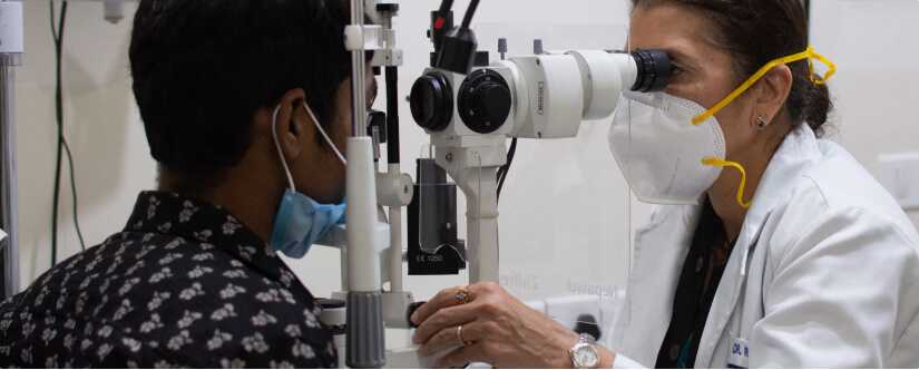
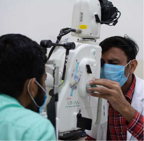
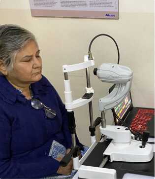
At Vision Plus Eye Centre we do a thorough clinical examination of your cornea, and if Keratoconus is suspected we have advanced machines that map the cornea and compare it to a normal cornea to flag any differences.
The methods include
- Retinoscopy- scissoring reflexes
Slit Lamp evaluation- conical shape, prominent nerves, corneal scarring - Keratometry – >48D
- OCT Pachymetry- Corneal thinning usually < 500um
- Pentacam- Most reliable technique to diagnose early keratoconus
The exact cause of Keratoconus are not fully understood yet, research is still being conducted.
- Frequent eye rubbing
- Eye allergies
- Family History
- Downs Syndrome, Ehler Danlos, Retinitis pigmentosa
- Environmental factors
- Asthma
- Blurred vision
- Ghosting of images
- Frequent change of glasses number
- Light sensitivity and glare
- Discomfort with glasses
At Vision Plus Eye Centre we do a thorough clinical examination of your cornea, and if Keratoconus is suspected we have advanced machines that map the cornea and compare it to a normal cornea to flag any differences.
The methods include
- Retinoscopy- scissoring reflexes
Slit Lamp evaluation- conical shape, prominent nerves, corneal scarring - Keratometry – >48D
- OCT Pachymetry- Corneal thinning usually < 500um
- Pentacam- Most reliable technique to diagnose early keratoconus
The exact cause of Keratoconus are not fully understood yet, research is still being conducted.
- Frequent eye rubbing
- Eye allergies
- Family History
- Downs Syndrome, Ehler Danlos, Retinitis pigmentosa
- Environmental factors
- Asthma
What are the treatment options for keratoconus.
These can help correct vision in the early stages
Special types of contact lenses like Rose K, piggy back, Miniscleral, hybrid lenses help to improve the vision quality. These are a simple way to correct the vision in mild to moderate cases.
This is a new technique to halt the progression of Keratoconus. Using Riboflavin dye and UV light to form cross linkages between the collagen fibrils within the corneal stroma. This helps stiffen the corneal tissue, strengthen it and reduce the ectasia or bulging. It is a daycare procedure which takes about 30 minutes. The outer layer of epithelium is first gently removed, then riboflavin drops are instilled and the cornea is exposed to a special UV light. A bandage contact lens is placed for a few days. Recovery can take 1 to 3 weeks and vision slowly improves.
At VPEC, we have expertise in handling advanced cases with thin corneas <400u
Patients with very steep and irregular corneas have poor quality of vision which cannot be corrected by glasses or contact lenses. INTACS or Intracorneal ring segments are customised semicircular rings made of PMMA plastic that are inserted into a channel made by a Femtosecond laser within the cornea. The size and number of the rings depends upon the extent of Keratoconus. They regularise and flatten the conical cornea making it more spherical and thus improving vision.It can be combined with C3R to prevent progression.
To improve quality of vision, Topo Guided PRK can be done alone or combined with Collage cross linking. In this Excimer laser is used to reshape the cornea and regularise the asymmetry and aberrations. It helps to improve the best corrected vision.
For patients who want freedom from glasses and contact lenses, Implantable contact lenses are an excellent option. They are a permanent solution unlike contact lenses and are made of a special thin plastic customised to the patients power and dimensions of their eye.The lens is implanted through a tiny incision in front of the natural lens. Recovery is rapid and the patient regains good quality of vision. For cases with high astigmatism, Toric Phakic IOLs are available. Keratoconus should be stable for at least 1 year before implanting these custom made ICLs.
In advanced Keratoconus where the cornea is extremely thin, conical and scarred, none of the conservative measures to correct vision help. As a last resort, cornea transplant is required. In this the patient’s diseased cornea is replaced with a donor cornea. It may be a full thickness tissue transplant called Penetrating Keratoplasty (PK) or a partial replacement of the front layers called Deep Anterior Lamellar Keratoplasty (DALK). The donor cornea is kept in place with stitches and requires a close follow up with the Cornea specialist. VPEC is recognised as a Cornea Transplant Centre by NOTTO. We have an experienced and dedicated team of Cornea specialists who carefully follow up the patients as there are risks of rejection and inflammation.
CORNEAL EDEMA
The Cornea is the transparent dome shaped outer layer of the eye that helps to focus light onto the Retina. In order to function optimally the cornea must be clear. When the cornea gets swollen due to excess fluid within its layers, the condition is called Corneal Edema. It results in decreased and cloudy vision.
What are the causes?
The innermost layer called the Endothelium acts like a pump to remove excess fluid from within the cornea and thus ensure it remains transparent. Any condition that affects the functioning of the endothelium can result in corneal edema.
- Cataract surgery
- Fuch’s Endothelial Dystrophy- more in females, above 50 years
- Viral Endotheliitis- Herpes virus
- Contact lens overuse/ sleeping with lenses on
- Inflammation of the cornea- due to Rheumatoid arthritis, keratitis
- Preservatives like Benzalkonium
- Drugs- Dorzolamide
How can it be treated?
- Cloudy vision especially on waking up
- Decreased vision
- Painful blisters
- Foreign body sensation
- Colour halos around lights
What are the Symptoms?
- Medications like saline drops to draw out the fluid from the cornea
- Steroid drops to reduce the inflammation
- Bandage contact lenses to reduce the pain from the blisters
- Antivirals
- Endothelial Keratoplasty- Replacing the diseased endothelium with a donor graft
- Penetrating Keratoplasty- Transplanting the entire cornea with a donor cornea
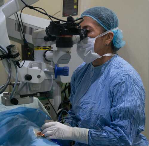
CORNEAL TRANSPLANT SURGERY/ KERATOPLASTY
The Cornea is an extremely delicate structure. Disease or injury can cause permanent damage, scarring and impair its functioning. Blindness due to corneal disease is the 4th most common cause of global blindness. Replacement of the diseased or opaque cornea by a part or whole of a healthy donor cornea is called Corneal Transplantation or Keratoplasty.
When a deceased person’s eyes are donated, the eye bank preserves the cornea such that it remains vital. The donated cornea which is transplanted to the recipients eyes is called Corneal graft.
Vision Plus Eye Centre is registered with NOTTO (National Organ & Tissue Transplant Organisation) and our cornea specialists are well experienced in all types of Cornea transplant surgery.
Indications for Cornea Transplant surgery
- Corneal scar/ opacity significantly affecting vision
- Keratoconus or thinning of the cornea
- Hereditary corneal dystrophy
- Bullous keratopathy after eye surgery
- Corneal infection not responding to medical therapy
Types of Cornea Transplant surgery
- Penetration Keratoplasty(PK) – Full thickness of the cornea is replaced. A trephine is used to remove a button of the damaged cornea and an appropriate size of donor cornea is then stitched in place. There is risk of corneal rejection and graft failure. Long term use of medications are required to prevent this.
- Deep Anterior Lamellar Keratoplasty (DALK) – The anterior diseased layers are replaced by donor tissue leaving behind the healthy endothelium of the recipient.
- Endothelial Keratoplasty (DSAEK /DMEK) – Only the damaged endothelium which is the thin innermost layer is replaced by the donor tissue. This has lesser chances of rejection and early recovery
Cost of Cornea transplant surgery in Delhi/NCR?
Vision Plus Eye Centre is one of the most affordable eye hospitals for surgery. Our Cornea specialists are highly experienced and give personalised support as cornea transplant surgery requires regular check up. After thorough evaluation, our expert doctors will recommend type of cornea transplant surgery and give the most reasonable estimate
PTERYGIUM
OCULAR HERPES
Herpes virus can affect the eyes causing serious infection of the cornea. There are 2 types of Herpes virus- Herpes simplex and Herpes varicella zoster. If detected and treated timely, complications like corneal scar can be reduced. Treatment is by antiviral drugs. Since virus infections can be recurrent, sometimes long term oral antiviral therapy is advised.